Hroscopic Medial Gutter Knee

The Lateral Gutter Drive Through Sign An Arthroscopic Indicator Of Acute Femoral Avulsion Of The Popliteus Tendon In Knee Joints Sciencedirect

The Knee Musculoskeletal Key

Meniscus Tears Understanding A Common Knee Problem Uvm Medical Center Blog
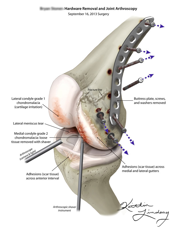
Knee Arthroscopy Hardware Removal Kaitlin Lindsay
Https Www Ajronline Org Doi Pdf 10 2214 Ajr 177 1 1770221

If You Have Osteoarthritis You Need Massage Massage Magazine Knee Osteoarthritis Osteoarthritis Knee Arthritis

From the medial compartment the lateral aspect of the medial femoral condyle and meniscus is assessed.
Hroscopic medial gutter knee.

Step By Step Arthroscopic Assessment Of The Anterolateral Ligament Of The Knee Using Anatomic Landmarks Sciencedirect

Figure 1 From Arthroscopic Treatment Of The Arthrofibrotic Knee Semantic Scholar
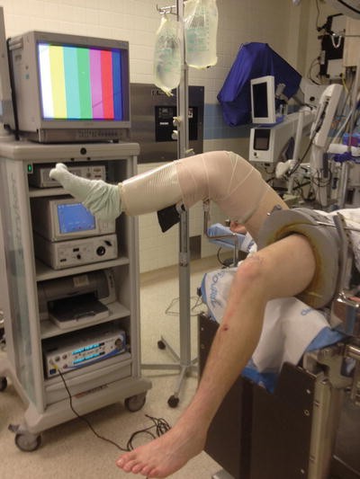
Knee Arthroscopy Setup Diagnosis Portals And Approaches Musculoskeletal Key

Can Knee Braces Help You Recover From A Torn Meniscus Injury Mcdavid
Knee Arthroscopy Orthobullets Orthobullets Knee Arthroscopy

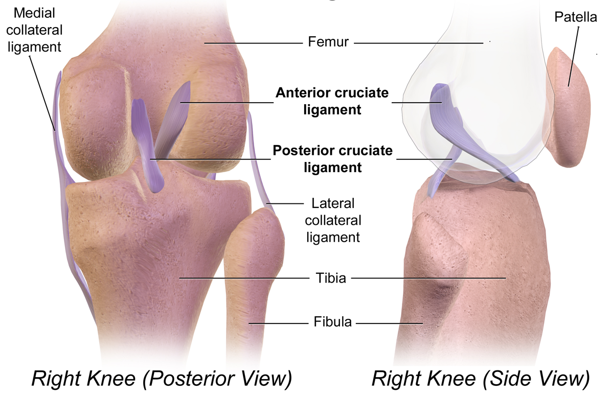
Anatomy Of The Knee Bones Muscles Arteries Veins Nerves Anatomy Of The Knee Joints Anatomy Muscle Anatomy

Lateral And Posterolateral Corner Injuries Of The Knee Musculoskeletal Key

Complete Arthroscopic Synovectomy In Management Of Recalcitrant Septic Arthritis Of The Knee Joint Sciencedirect

Knee Diagnostic Arthroscopy In Left Knee After Gunshot Wound The Download Scientific Diagram
Arif Arthroscopic Reduction And Internal Fixation Around The Elbow Springerlink
Diagnostic Knee Arthroscopy And Arthroscopic Anatomy Springerlink

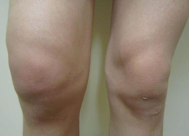
Case Study Management Of Medial Meniscal Tear And Patellar Osteochondral Damage In A 60 Year Old Female

Perfect Fit Health Shoulder Posture Exercise Personal Training

The Lateral Gutter Drive Through Sign Revisited A Cadaveric Study Exploring Its Real Mechanism Based On The Individual Posterolateral Structure Of Knee Joints Springerlink
Https Www Jorgechahlamd Com Wp Content Uploads 2019 12 215 Root Pdf
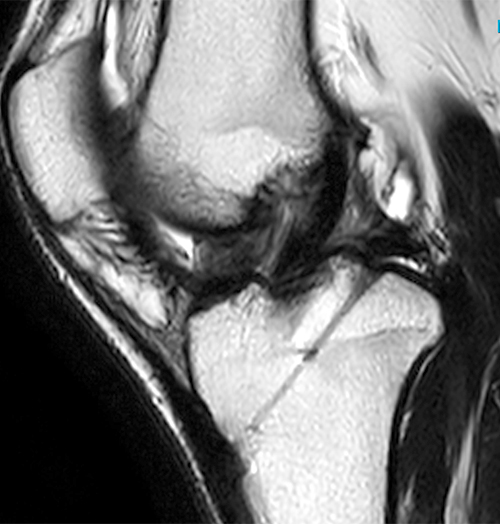
Arthrofibrosis Of The Knee Radsource
Http Www Remedypublications Com Open Access Pthe Quotfourth Compartmentquot Of The Knee A New Paradigmp 2147 Pdf

Single Portal Knee Arthroscopy 2015 Technique Update Sciencedirect
Https Encrypted Tbn0 Gstatic Com Images Q Tbn 3aand9gcq40mjpiinu01yxzk22jpk0dadzevvvgs2zvl2qnnvpybuvalvv Usqp Cau
Musculoskeletal

Pdf Popliteomeniscal Fascicle Tears Causing Symptomatic Lateral Compartment Knee Pain Diagnosis By The Figure 4 Test And Treatment By Open Repair

Arthroscopic Techniques To Enhance Meniscus Visualization Sciencedirect
Examination Of The Knee Joint Teachmesurgery
Https Tricemedical Com Wp Content Uploads Technique Paper Mcmillan Arthroscopy Techniques Pdf
Source : pinterest.com
